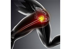
Knee
Dr. Burns specializes in treatment of complex sports injuries of the knee. This includes meniscal tears, cartilage injuries, ACL tears, PCL tears, MCL/LCL tears, patellar instability, and knee malalignment. We perform complex ligament reconstructions, comprehensive meniscal treatment from debridement to meniscal transplants, and cartilage replacement procedures such as OATs allograft and MACI.
Please see the links below for information on knee anatomy, injuries and surgical treatments. Please also review the patient info tab above to find new patient forms, preoperative instructions, postoperative instructions, and physical therapy guidelines for these procedures.
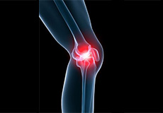
Knee Anatomy
The knee is a complex joint made up of different structures including bones, tendons, ligaments and muscles. They all work together to maintain normal function and provide stability to the knee during movement.
Having a well-functioning healthy knee is essential for our mobility and ability to participate in various activities. Understanding the anatomy of the knee enhances your ability to discuss and choose the right treatment procedure for knee problems with your doctor.
Bones of the Knee
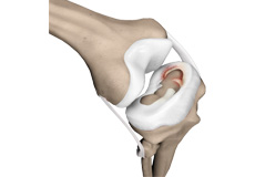
The knee is a hinge joint made up of two bones, the thighbone (femur) and the shinbone (tibia). There are two round knobs at the end of the femur called femoral condyles which articulate with the flat surface of the tibia called the tibial plateau. The tibia plateau on the inside of the leg is called the medial tibial plateau, and on the outside of the leg it is called the lateral tibial plateau.
The two femoral condyles form a groove on the front (anterior) side of the knee called the patellofemoral groove. A small bone called the patella sits in this groove and forms the kneecap. It acts as a shield and protects the knee joint from direct trauma.
A fourth bone called the fibula is the other bone of the lower leg. This forms a small joint with the tibia. This joint has very little movement and is not considered a part of the main joint of the knee.
Articular Cartilage and Menisci of the Knee
Movement of the bones causes friction between the articulating surfaces. To reduce this friction, all articulating surfaces involved in movement are covered with a white, shiny, slippery layer called articular cartilage. The articulating surface of the femoral condyles, tibial plateaus and the back of the patella are covered with this cartilage. The cartilage provides a smooth surface that facilitates easy movement.
To further reduce friction between the articulating surfaces of the bones, the knee joint is lined by a synovial membrane which produces a thick clear fluid called synovial fluid. This fluid lubricates and nourishes the cartilage and bones inside the joint capsule.
Within the knee joint, between the femur and tibia, there are two C shaped cartilaginous structures called menisci. Menisci function to provide stability to the knee by spreading the weight of the upper body across the whole surface of the tibial plateau. The menisci help in load- bearing by preventing the weight from concentrating onto a small area, which could damage the articular cartilage. The menisci also act as a cushion between the femur and tibia by absorbing the shock produced by activities such as walking, running and jumping.
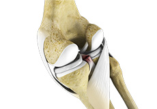
Ligaments of the Knee
Ligaments are tough bands of tissue that connect one bone to another bone. The ligaments of the knee function to stabilize the knee joint. There are two important groups of ligaments that hold the bones of the knee joint together, collateral ligaments and the cruciate ligament.
Collateral ligaments are present on either side of the knee. They function to prevent the knee from moving too far during side to side motion. The collateral ligament on the inside is called the medial collateral ligament (MCL) and the collateral ligament on the outside is called the lateral collateral ligament (LCL).
Cruciate ligaments, present inside the knee joint, control the back-and-forth motion of the knee. The cruciate ligament in the front of the knee is called anterior cruciate ligament or ACL and the cruciate ligament in the back of the knee is called posterior cruciate ligament or PCL.
Muscles of the Knee
Muscles: There are two major muscles, the quadriceps and the hamstrings, which enable movement of the knee joint. The quadriceps muscles are in the front of the thigh. When the quadriceps muscles contract, the knee straightens. The hamstrings are in the back of the thigh. When the hamstring muscles contract, the knee bends.
Tendons of the Knee
Tendons are structures that attach muscles to the bone. The quadriceps muscles of the knee meet just above the patella and attach to it through a tendon called the quadriceps tendon. The patella further attaches to the tibia through a tendon called the patella tendon. The quadriceps muscle, quadriceps tendon and patellar tendon all work together to straighten the knee. Similarly, the hamstring muscles at the back of the leg are attached to the knee joint with the hamstring tendon.
Conditions
Knee Pain
The knee is one of the largest joints in the body, formed by the lower end of the femur, upper end of the tibia and the patella or kneecap. Several ligaments and muscles attach to the bones of the knee joint to maintain normal motion of the joint.
Knee Injury
The knee is a hinge joint made up of two bones, the thighbone (femur) and the shinbone (tibia), which articulate with each other. A groove on the front side of the knee called the patellofemoral groove sits a small bone called the patella or kneecap.
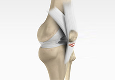
Unstable Knee
The knee joint is one of the largest joints in the body. This highly complex joint has several tissues supporting and stabilizing its movement

Ligament Injuries
The knee is a complex joint which consists of bone, cartilage, ligaments and tendons that make joint movements easy and at the same time more susceptible to various kinds of injuries.
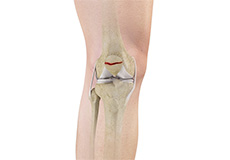
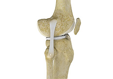
Anterior Cruciate Ligament (ACL) Tears
The anterior cruciate ligament, or ACL, is one of the major ligaments of the knee that is in the middle of the knee and runs from the femur (thighbone) to the tibia (shinbone). It prevents the tibia from sliding out in front of the femur. Together with posterior cruciate ligament (PCL) it provides rotational stability to the knee.
Medial Collateral Ligament (MCL)Tears
The medial collateral ligament (MCL) is the ligament that is located on the inner part of the knee joint. It runs from the femur (thighbone) to the top of the tibia (shinbone) and helps in stabilizing the knee.
Posterior Cruciate Ligament (PCL) Injury
Posterior cruciate ligament (PCL), one of four major ligaments of the knee are situated at the back of the knee. It connects the thighbone (femur) to the shinbone (tibia). The PCL limits the backward motion of the shinbone.

Lateral Collateral Ligament (LCL) Injury
Lateral collateral ligament (LCL) is a thin set of tissues present on the outer side of the knee, connecting the thighbone (femur) to the fibula (side bone of lower leg). It provides stability as well as limits the sidewise rotation of the knee. Tear or injury of theLCL may cause instability of the knee that can be either reconstructed or repaired to regain the strength and movement of the knee.
Multiligament Instability
The knee is a complex joint of the body which is vital for movement. The four major ligaments of the knee are anterior cruciate ligament, posterior cruciate ligament, medial collateral ligament and lateral collateral ligament. They play an important role in maintaining the stability of the knee. An injury resulting in tear of one or more ligaments of the knee thus affects knee stability.
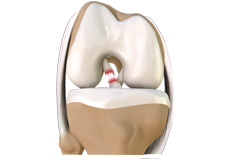
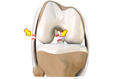
Meniscal Tears
Meniscus tear is the commonest knee injury in athletes, especially those involved in contact sports. A sudden bend or twist in your knee causes the meniscus to tear. This is a traumatic meniscus tear. Elderly people are more prone to degenerative meniscal tears as the cartilage wears out and weakens with age.
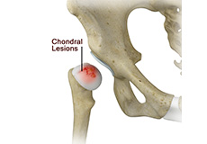
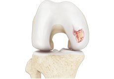
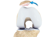
Chondral (Articular Cartilage Defects)
Articular or hyaline cartilage is the tissue lining the surface of the two bones in the knee joint. Cartilage helps the bones move smoothly against each other and can withstand the weight of the body during activities such as running and jumping.
Knee Arthritis
Arthritis is a general term covering numerous conditions where the joint surface or cartilage wears out. The joint surface is covered by a smooth articular surface that allows pain free movement in the joint. This surface can wear out for several reasons; often the definite cause is not known.
Patellar Dislocation/Patellofemoral Dislocation
Patella (kneecap) is a protective bone attached to the quadriceps muscles of the thigh by quadriceps tendon. Patella attaches with the femur bone and forms a patellofemoral joint. Patella is protected by a ligament which secures the kneecap from gliding out and is called as medial patellofemoral ligament (MPFL).
Patellar Instability
Patellar (kneecap) instability results from one or more dislocations or partial dislocations (subluxations). Patella is the small piece of bone in front of the knee that slides up and down the femoral groove (groove in the femur bone) during bending and stretching movements.
Patella Fracture
The kneecap or patella is the largest sesamoid bone in the body and one of the components of the knee joint, present at the front of the knee. The undersurface of the kneecap and the lower end of the femur are coated with articular cartilage, which helps in smooth movement of the knee joint. The kneecap protects the knee and provides attachment to various muscle groups of the thigh and leg. Fracture of kneecap is rare and is more common in adult males.
Non-Surgical Treatments

Pharmacological
The knee is a complex joint which consists of bone, cartilage, ligaments and tendons that make joint movements easy and at the same time more susceptible to various kinds of injuries. Knee problems may arise if any of these structures get injured by overuse or suddenly during sports activities.
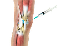
Viscosupplementation
Viscosupplementation refers to the injection of a hyaluronan preparation into the joint. Hyaluronan is a natural substance present in the joint fluid that assists in lubrication. It allows smooth movement of the cartilage covered articulating surfaces of the joint.
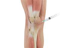
Cortisone Injection
Cortisone is a corticosteroid released by the adrenal gland in response to stress and is a potent anti-inflammatory agent.
Physical Therapy
Physiotherapy or physical therapy is an exercise program that helps you to improve movement, relieve pain, encourage blood flow for faster healing, and restore your physical function and fitness level. It can be prescribed as an individual treatment program or combined with other treatments. It involves a combination of education, manual therapy, exercises and techniques such as water, heat, cold, electrical stimulation and ultrasound.
Surgical Treatment
Knee Arthroscopy
The knee joint is one of the most complex joints of the body. The lower end of the thighbone (femur) meets the upper end of the shinbone (tibia) at the knee joint. A small bone called the patella (kneecap) rests on a groove on the front side of the femoral end. A bone of the lower leg (fibula) forms a joint with the shinbone.
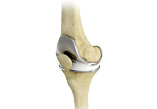
Total Knee Replacement
Total knee replacement, also called total knee arthroplasty, is a surgical procedure in which the worn out or damaged surfaces of the knee joint are removed and replaced with an artificial prosthesis.
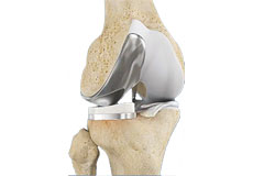
Partial Knee Replacement
Unicompartmental knee replacement is a minimally invasive surgery in which only the damaged compartment of the knee is replaced with an implant. It is also called a partial knee replacement.
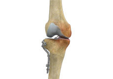
High Tibial Osteotomy
High tibial osteotomy is a surgical procedure performed to relieve pressure on the damaged site of an arthritic knee joint. It is usually performed in arthritic conditions affecting only one side of your knee and the aim is to take pressure off the damaged area and shift it to the other side of your knee with healthy cartilage.
Tibial Tubercle Osteotomy
Tibial tubercle osteotomy is a surgical procedure which is performed along with other procedures to treat patellar instability, patellofemoral pain, and osteoarthritis. Tibial tubercle transfer technique involves realignment of the tibial tubercle (a bump in the front of the shinbone) such that the kneecap (patella) traverses in the center of the femoral groove.
Quadriceps Tendon Repair
Quadriceps tendon is a thick tissue located at the top of the kneecap. The quadriceps tendon works together with the quadriceps muscles to allow us to straighten our leg. The quadriceps muscles are the muscles located in front of the thigh.
Patellar Tendon Repair
Patella tendon rupture is the rupture of the tendon that connects the patella (kneecap) to the top portion of the tibia (shinbone). The patellar tendon works together with the quadriceps muscle and the quadriceps tendon to allow your knee to straighten out.
MACI® Autologous Chondrocyte Implantation
Matrix-induced autologous chondrocyte implantation (MACI) is a procedure to treat articular cartilage defects of the knee. This procedure is effective for treating small areas of cartilage damage that causes pain and swelling and restricts the range of motion. Autologous chondrocyte implantation is not indicated for those patients who have advanced arthritis of the knee.
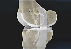
ACL Reconstruction
The anterior cruciate ligament is one of the major stabilizing ligaments in the knee. It is a strong rope like structure located in the center of the knee running from the femur to the tibia. When this ligament tears unfortunately, it does not heal and often leads to the feeling of instability in the knee.
ACL Meniscus Repair
Coming soon
ACL Reconstruction (Bone - Patellar Tendon – Bone)
Anterior cruciate ligament (ACL) reconstruction (bone - patellar tendon – bone) is a surgical procedure that replaces the injured ACL with part of the patellar tendon and two plugs of bone at its ends.
ACL Revision Reconstruction
Coming soon
ACL Reconstruction – Hamstring Tendon
The anterior cruciate ligament is one of the major stabilizing ligaments in the knee. It is a strong rope like structure located in the center of the knee running from the femur to the tibia. When this ligament tears unfortunately, it does not heal and often leads to the feeling of instability in the knee.
Medial Patellofemoral Ligament Reconstruction
Medial patellofemoral ligament reconstruction is a surgical procedure indicated in patients with more severe patellar instability. Medial patellofemoral ligament is a band of tissue that extends from the femoral medial epicondyle to the superior aspect of the patella.
Meniscal Repair
A meniscal tear is a tear that occurs in the cartilage of the knee. The meniscus is a small, "C" shaped piece of cartilage in the knee joint. Each knee has two menisci, the medial meniscus on the inner aspect of the knee and the lateral meniscus on the outer aspect of the knee. The medial and lateral menisci act as a cushion between the thigh bone (femur) and shin bone (tibia).
Meniscal Allograft Transplant
Meniscal transplantation is a surgical procedure to replace the damaged meniscus of the knee with healthy cartilage from a donor.
The meniscus is a C-shaped cartilage ring that acts like a cushion between the shinbone and the thighbone. Each of your knees has two menisci - one on the inside (medial aspect) and the other on the outside (lateral aspect) of your knee. Apart from the cushioning effect, the menisci also provide stability to the knee.
Posterior Cruciate Ligament (PCL) Reconstruction
Posterior cruciate ligament (PCL), one of four major ligaments of the knee are situated at the back of the knee. It connects the thighbone (femur) to the shinbone (tibia). The PCL limits the backward motion of the shinbone.
Lateral Collateral Ligament (LCL) Reconstruction
Lateral collateral ligament (LCL) is a thin set of tissues present on the outer side of the knee, connecting the thighbone (femur) to the fibula (side bone of lower leg). It provides stability as well as limits the sidewise rotation of the knee. Tear or injury of LCL may cause instability of the knee that can be either reconstructed or repaired to regain the strength and movement of the knee.
Cartilage Replacement
Cartilage replacement is a surgical procedure performed to replace the worn-out cartilage with the new cartilage. It is usually performed to treat patients with small areas of cartilage damage usually caused by sports or traumatic injuries. It is not indicated for those patients who have advanced arthritis of knee.
Cartilage Repair & Transplantation
Articular Cartilage is the white tissue lining the end of bones where these bones connect to form joints. Cartilage acts as cushioning material and helps in smooth gliding of bones during movement. An injury to the joint may damage this cartilage which cannot repair on its own. Cartilage can be damaged with increasing age, normal wear and tear, or trauma. Damaged cartilage cannot cushion the joints during movement and the joints may rub over each other causing severe pain and inflammation.
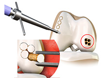
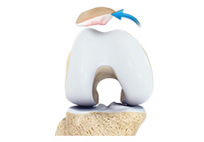
OATS
OATS is “osteochondral autograft transfer system”. It is one of the two types of cartilage transfer procedures and the other procedure is “Mosaicplasty”. Cartilage transfer procedures involve moving healthy cartilage from a non-weight bearing area of the knee to a damaged area of the cartilage in the knee.