
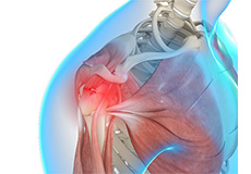
Shoulder & Elbow
Dr. Burns specializes in treatment of shoulder injuries including arthroscopic and open procedures. Arthroscopic treatment focuses on labral tears, shoulder instability, biceps pathology, rotator cuff tears, impingement syndrome, and acromioclavicular arthritis. Open shoulder reconstructions are necessary for some shoulder fractures, shoulder separations, revision instability operations, and shoulder replacements. We utilize the latest technology in performing anatomic and reverse shoulder replacements. Recent innovations in implants and OR technology allow for use of “stemless” shoulder replacements and the use of computer assisted implant positioning which has be shown to improve our ability to position the replacement components.
Please see the links below for information on shoulder and elbow anatomy, injuries and surgical treatments. Please also review the patient info tab above to find new patient forms, preoperative instructions, postoperative instructions, and physical therapy guidelines for these procedures.
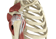
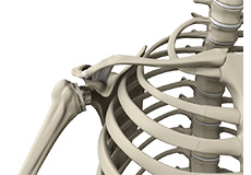
Shoulder Anatomy
The shoulder is the most flexible joint in the body enabling a wide range of movements including, forward flexion, abduction, adduction, external rotation, internal rotation, and 360-degree circumduction.
Thus, the shoulder joint is considered the most insecure joint of the body but the support of ligaments, muscles and tendons function to provide the required stability.
Bones of the Shoulder
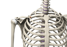
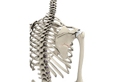
The shoulder is a ball and socket joint made up of three bones, namely the humerus, scapula, and clavicle.
The end of the humerus or upper arm bone forms the ball of the shoulder joint. An irregular shallow cavity in the scapula called the glenoid cavity forms the socket for the head of the humerus to fit in. The two bones together form the glenohumeral joint, which is the main joint of the shoulder.
The scapula is a flat triangular-shaped bone that forms the shoulder blade. It serves as the site of attachment for most of the muscles that provide movement and stability to the joint. The scapula has four bony processes - acromion, spine, coracoid and glenoid cavity. The Acromion and coracoid process serve as places for attachment of the ligaments and tendons.
The clavicle bone or collarbone is an S-shaped bone that connects the scapula to the sternum or breastbone. It forms two joints: the acromioclavicular joint, where it articulates with the acromion process of the scapula, and the sternoclavicular joint where it articulates with the sternum or breast bone. The clavicle also forms a protective covering for important nerves and blood vessels that pass under it from the spine to the arms.
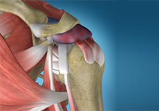
Soft Tissues of the Shoulder
The ends of all articulating bones are covered by smooth tissue called articular cartilage which allows the bones to slide over each other without friction enabling smooth movement. Articular cartilage reduces pressure and acts as a shock absorber during movement of the shoulder bones.
Extra stability to the glenohumeral joint is provided by the glenoid labrum, a ring of fibrous cartilage that surrounds the glenoid cavity. The glenoid labrum increases the depth and surface area of the glenoid cavity to provide a more secure fit for the half-spherical head of the humerus.
Ligaments of the Shoulder
Ligaments are the thick strands of fibers that connect one bone to another. The ligaments of the shoulder joint include
- Coraco-clavicular ligaments: These ligaments connect the collarbone to the shoulder blade at the coracoid process
- Acromio-clavicular ligament: This connects the collarbone to the shoulder blade at the acromion process
- Coraco-acromial ligament: It connects the acromion process to the coracoid process
- Glenohumeral ligaments: A group of 3 ligaments that form a capsule around the shoulder joint, and connect the head of the arm bone to the glenoid cavity of the shoulder blade. The capsule forms a water-tight sac around the joint. Glenohumeral ligaments play a very important role in providing stability to the otherwise unstable shoulder joint by preventing dislocation.
Muscles of the Shoulder
The rotator cuff is the main group of muscles in the shoulder joint and is comprised of 4 muscles. The rotator cuff forms a sleeve around the humeral head and glenoid cavity, providing additional stability to the shoulder joint while enabling a wide range of mobility.
The deltoid muscle forms the outer layer of the rotator cuff and is the largest and strongest muscle of the shoulder joint.
Tendons of the Shoulder
Tendons are strong tissues that join muscle to bone allowing the muscle to control the movement of the bone or joint. Two important group of tendons in the shoulder joint are the biceps tendons and rotator cuff tendons.
Bicep tendons are the two tendons that join the bicep muscle of the upper arm to the shoulder. They are referred to as the long head and short head of the bicep.
Rotator cuff tendons are a group of four tendons that join the head of the humerus to the deeper muscles of the rotator cuff. These tendons provide more stability and mobility to the shoulder joint.
Nerves of the Shoulder
Nerves carry messages from the brain to muscles to direct movement (motor nerves) and send information about different sensations such as touch, temperature and pain from the muscles back to the brain (sensory nerves). The nerves of the arm pass through the shoulder joint from the neck.
These nerves form a bundle at the region of the shoulder called the brachial plexus. The main nerves of the brachial plexus are the musculocutaneous, axillary, radial, ulnar and median nerves.
Blood vessels of the Shoulder
Blood vessels travel along with the nerves to supply blood to the arms. Oxygenated blood is supplied to the shoulder region by the subclavian artery that runs below the collarbone. As it enters the region of the armpit, it is called the axillary artery and further down the arm, it is called the brachial artery. The main veins carrying de-oxygenated blood back to the heart for purification include:
- Axillary vein: this vein drains into the subclavian vein
- Cephalic vein: this vein is found in the upper arm and branches at the elbow into the forearm region. It drains into the axillary vein.
- Basilic vein: this vein runs opposite the cephalic vein, near the triceps muscle. It drains into the axillary vein.
Elbow Anatomy
The elbow is a complex joint formed by the articulation of three bones –the humerus, radius and ulna. The elbow joint helps in bending or straightening of the arm to 180 degrees and assists in lifting or moving objects.
The bones of the elbow are supported by
- Ligaments and tendons
- Muscles
- Nerves
- Blood vessels
Bones and Joints of the elbow joint:
The elbow joint is formed at the junction of three bones:
- The Humerus (upper arm bone) forms the upper portion of the joint. The lower end of the humerus divides in to two bony protrusions known as the medial and lateral epicondyles which can be felt on either side of the elbow joint.
- The Ulna is the larger bone of the forearm located on the inner surface of the joint. The curved shape of the ulna articulates with the humerus.
- The Radius is the smaller bone of the forearm situated on the outer surface of the joint. The head of the radius is circular and hollow which allows movement with the humerus. The connection between the ulna and radius helps the forearm to rotate.
The elbow consists of three joints from articulation of the three bones namely:
- Humero-ulnar joint is formed between the humerus and ulna and allows flexion and extension of the arm.
- Humero-radial joint is formed between the radius and humerus, and allows movements like flexion, extension, supination and pronation.
- Radio-ulnar joint is formed between ulna and radius bones, and allows rotation of the lower arm.
Articular cartilage lines the articulating regions of the humerus, radius and ulna. It is a thin, tough, flexible, and slippery surface that acts as a shock absorber and cushion to reduce friction between the bones. The cartilage is lubricated by synovial fluid, which further enables the smooth movement of the bones.
Muscles of the Elbow Joint
There are several muscles extending across the elbow joint that help in various movements. These include the following:
- Biceps brachii: upper arm muscle enabling flexion of the arm
- Triceps brachii: muscle in the back of the upper arm that extends the arm and fixes the elbow during fine movements
- Brachialis: upper arm muscle beneath the biceps which flexes the elbow towards the body
- Brachioradialis: forearm muscle that flexes, straightens and pulls the arm at the elbow
- Pronator teres: this muscle extends from the humeral head, across the elbow, and towards the ulna, and helps to turn the palm facing backward
- Extensor carpi radialis brevis: forearm muscle that helps in movement of the hand
- Extensor digitorum: forearm muscle that helps in movement of the fingers
Elbow joint ligaments and tendons:
The elbow joint is supported by ligaments and tendons, which provide stability to the joint.
Ligaments are a group of firm tissues that connect bones to other bones. The most important ligaments of the elbow joint are the:
- Medial or ulnar collateral ligament: comprised of triangular bands of tissue on the inner side of the elbow joint.
- Lateral or radial collateral ligament: a thin band of tissue on the outer side of the elbow joint.
Together, the medial and lateral ligaments are the main source of stability and hold the humerus and ulna tightly in place during movement of the arm.
- Annular ligament: These are a group of fibers that surrounds the radial head, and holds the ulna and radius tightly in place during movement of the arm.
The ligaments around a joint combine to form a joint capsule that contains synovial fluid.
Any injury to these ligaments can lead to instability of the elbow joint.
Tendons are bands of connective tissue fibers that connect muscle to bone. The various tendons which surround the elbow joint include:
- Biceps tendon: attaches the biceps muscle to the radius, allowing the elbow to bend
- Triceps tendon: attaches the triceps muscle to the ulna, allowing the elbow to straighten
Nerves of the elbow joint:
The main nerves of the elbow joint are the ulnar, radial and median nerves. These nerves transfer signals from the brain to the muscles that aid in elbow movements. They also carry the sensory signals like touch, pain, and temperature back to the brain.
Any injury or damage to these nerves causes pain, weakness or joint instability.
Blood vessels:
Arteries are blood vessels that carry oxygen-pure blood from the heart to the hand. The main artery of the elbow is the brachial artery that travels across the inside of the elbow and divides into two small branches below the elbow to form the ulnar and the radial artery.
Conditions
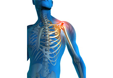
Shoulder Pain
Pain in the shoulder suggests a shoulder injury which is more common in athletes participating in sports such as swimming, tennis, pitching and weightlifting. The injuries are caused due to the over usage or repetitive motion of the arms.
Rotator Cuff Tear
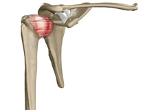
Rotator cuff is the group of tendons in the shoulder joint providing support and enabling wider range of motion. Major injury to these tendons may result in tear of these tendons and the condition is called as rotator cuff tear. It is one of the most common causes of shoulder pain in middle aged adults and older individuals.

Subluxation
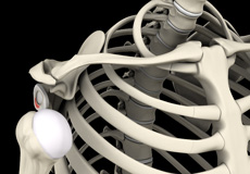
The shoulder is a highly mobile ball and socket joint. The ball of the upper arm bone (humerus) is held in place at the socket (glenoid) of the shoulder blade (scapula) by a group of ligaments. A partial dislocation of the shoulder joint is termed as a subluxation. This means the ball has partially moved out of the glenoid as opposed to a dislocation, where the ball completely moves out of the glenoid.
Shoulder Impingement
Shoulder impingement is the condition of inflammation of the tendons of the shoulder joint. It is one of the most common causes of pain in the adult shoulder. The shoulder is a 'ball-and-socket' joint. A ‘ball' at the top of the upper arm bone, humerus, fits neatly into a 'socket’, called the glenoid, which is part of the shoulder blade, scapula. Shoulder impingement is also called as swimmer’s shoulder, tennis shoulder, or rotator cuff tendinitis.
SLAP Tears
The shoulder joint is a ball and socket joint. A 'ball' at the top of the upper arm bone (the humerus) fits neatly into a 'socket', called the glenoid, which is part of the shoulder blade (scapula). The term SLAP (superior –labrum anterior-posterior) lesion or SLAP tear refers to an injury of the superior labrum of the shoulder.
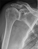
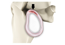
Arthritis of the Shoulder
The term arthritis literally means inflammation of a joint, but is generally used to describe any condition in which there is damage to the cartilage. Damage of the cartilage in the shoulder joint causes shoulder arthritis. Inflammation is the body's natural response to injury. The warning signs that inflammation presents are redness, swelling, heat and pain.
Frozen Shoulder
Frozen shoulder, also called adhesive capsulitis is a condition characterized by pain and loss of motion in shoulder joint. It is more common in older adults aged between 40 and 60 years and is more common in women than men.
Shoulder Instability
Shoulder instability is a chronic condition that causes frequent dislocations of the shoulder joint.
A dislocation occurs when the end of the humerus (the ball portion) partially or completely dislocates from the glenoid (the socket portion) of the shoulder. A partial dislocation is referred to as a subluxation whereas a complete separation is referred to as a dislocation.
Shoulder Labrum Tear
The shoulder joint is a “ball and socket” joint that enables the smooth gliding and thereby the movements of arms. However, it is inherently unstable because of the shallow socket. A soft rim of cartilage, the labrum lines the socket and deepens it so that it accommodates the head of the upper arm bone better.
Acromioclavicular (AC) Joint Injury
The acromioclavicular joint is part of the shoulder joint. It is formed by the union of the acromion, a bony process of the shoulder blade, and the outer end of the collar bone or clavicle. The joint is lined by cartilage that gradually wears with age as well as with repeated overhead or shoulder level activities such as basketball or bench pressing.

Shoulder Dislocation
Playing more overhead sports activities and repeated use of shoulder at workplace may lead to sliding of the upper arm bone, the ball portion, from the glenoid–the socket portion of the shoulder. The dislocation might be a partial dislocation (subluxation) or a complete dislocation causing pain and shoulder joint instability.
Rotator Cuff Tendonitis
The rotator cuff is a group of muscles and tendons that attach to the bones of the shoulder joint providing movement and stability to the shoulder. Inflammation of the rotator cuff tendons is called rotator cuff tendonitis or shoulder impingement.
Snapping Scapula Syndrome
Coming soon
Sternoclavicular Joint Injury
Coming soon
Scapular Winging
The scapula or shoulder blade is one of the 3 bones that make up the shoulder joint. It provides attachment for muscles that connect the upper arm to the body stabilizing the shoulder joint. These muscles act together to move the arm in different directions and hold the scapula in its correct position.
Bicep Tendon Rupture
The biceps muscle is present on the front side of your upper arm and functions to help you bend and rotate your arm.
The biceps tendon is a tough band of connective fibrous tissue that attaches your biceps muscle to the bones in your shoulder on one side and the elbow on the other side.
Distal Biceps Rupture
The biceps muscle is located in front of your upper arm. It helps in bending your elbow as well as in rotational movements of your forearm. Also, it helps to maintain stability in the shoulder joint. The biceps muscle has two tendons, one of which attaches it to the bone in the shoulder and the other attaches at the elbow. The biceps tendon at the elbow is called the distal biceps tendon and if there is a tear in this tendon, you will be unable to move your arm from the palm-down to palm-up position.
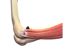
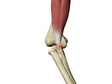
Tennis Elbow
Tennis elbow is the common name used for the elbow condition called lateral epicondylitis. It is an overuse injury that causes inflammation of the tendons that attach to the bony prominence on the outside of the elbow (lateral epicondyle). It is a painful condition occurring from repeated muscle contractions at the forearm that leads to inflammation and micro tears in the tendons that attach to the lateral epicondyle.
Pectoralis Major Rupture
The pectoralis major is a large muscle in the chest. It is used when you pull your shoulder across your chest. The pectoralis major originates from the clavicle (collar bone) as well as the sternum (breastbone) and is attached near the top of the upper arm bone (humerus).
Shoulder Trauma
Shoulder injuries most commonly occur in athletes participating in sports such as swimming, tennis, pitching, and weightlifting. The injuries are caused due to the over usage or repetitive motion of the arms.
Clavicle Fracture (Broken Collarbone)
Clavicle fracture, also called broken collarbone is a very common sports injury seen in people who are involved in contact sports such as football and martial arts as well as impact sports such as motor racing. A direct blow over the shoulder that may occur during a fall on an outstretched arm or a motor vehicle accident may cause the clavicle bone to break.
Fracture of the Shoulder Blade (Scapula)
The scapula (shoulder blade) is a flat, triangular bone providing attachment to the muscles of the back, neck, chest and arm. The scapula has a body, neck and spine portion.
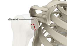
Glenoid Fractures
The glenoid is the socket that forms the ball and socket joint of the shoulder. Fractures of the glenoid are rare but can occur due to major trauma or during high-energy sports activities.
Non-Surgical Treatments
Ultrasound-guided Shoulder Injections
An ultrasound is a common imaging technique that employs high frequency sound waves to create images of organs and other internal structures of the body. These images provide valuable information of underlying pathology of the tissues and assists with diagnosis and planning the treatment of a condition. Ultrasound provides a clear view of the organs, tendons, muscles or joints and any associated disorders.
Surgical Treatment
AC Reconstruction
Coming soon
Biceps Tenodesis
The biceps is a large skeletal muscle of the upper arm that flexes the elbow to lift the forearm, and is also responsible for some shoulder movements. It is connected by tendons to bones in the shoulder and elbow on either side. Injury of the biceps tendon in the region of the shoulder can occur due to disease, overuse or repetitive overhead activity, leading to pain and weakness in front of the shoulder and down the upper arm.
Anterior Labral Repair
Coming soon
Posterior Labral Repair
Coming soon
Pectoralis Major Repair
Pectoralis major repair is surgery to repair the tendon of a large muscle in the chest called the pectoralis major. This muscle is used when you pull your shoulder across your chest. The pectoralis major originates from the clavicle (collar bone) as well as the sternum (breastbone) and is attached near the top of the upper arm bone (humerus).
Rotator Cuff Repair
The rotator cuff is a group of tendons in the shoulder joint providing support and enabling wider range of motion. Major injury to these tendons may result in tear of these tendons and the condition is called as rotator cuff tear.
SAD DCR Debridement
Coming soon
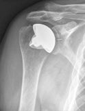
Total Shoulder Replacement
The shoulder is a highly movable body joint that allows various movements of the arm. It is a ball and socket joint, where the head of the humerus (upper arm bone) articulates with the socket of the scapula (shoulder blade) called the glenoid. The two articulating surfaces of the bones are covered with cartilage, which prevents friction between the moving bones.
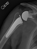
Reverse Shoulder Replacement
Reverse total shoulder replacement, is an advanced surgical technique specifically designed for rotator cuff tear arthropathy, a condition where the patient suffers from both shoulder arthritis and a rotator cuff tear.
Snapping Scapula Treatment
Coming soon
SLAP Repair
Your shoulder joint is a ball and socket joint made up of the upper arm bone, the shoulder blade and the collarbone. The head of the upper arm bone fits into the socket of the shoulder joint known as the glenoid cavity. The outer edge of the glenoid is surrounded by a strong fibrous tissue called the labrum.
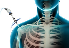
Shoulder Arthroscopy
Arthroscopy is a minimally invasive diagnostic and surgical procedure performed for joint problems. Shoulder arthroscopy is performed using a pencil-sized instrument called an Arthroscope. The arthroscope consists of a light system and camera to project images to a computer screen for your surgeon to view the surgical site. Arthroscopy is used to treat disease conditions and injuries involving the bones, cartilage, tendons, ligaments, and muscles of the shoulder joint.
Clavicle Fracture Repair
Clavicle fracture, also called broken collarbone, is a very common sports injury seen in people who are involved in contact sports such as football and martial arts as well as impact sports such as motor racing. A direct blow over the shoulder that may occur during a fall on an outstretched arm or a motor vehicle accident may cause the clavicle bone to break.
Latarjet Procedure
The shoulder joint provides a wide range of movement to the upper extremity, but overuse or trauma can cause instability to the joint. The Latarjet procedure is a surgical procedure performed to treat shoulder instability by relocating a piece of bone with an attached tendon to the shoulder joint.
Bankart Repair
The shoulder joint (glenohumeral joint) is a ball and socket joint, where the head of the upper arm bone (humerus) attaches to the shoulder socket (glenoid cavity). The shoulder socket is extremely shallow and therefore needs additional support to keep the shoulder bones from dislocating. The labrum, a cuff of cartilage that encircles the shoulder socket, helps serve this purpose by forming a cup for the humeral head to move within. It provides stability to the joint, enabling a wide range of movements.
Superior Capsular Reconstruction
The shoulder joint is stabilized by the joint capsule and rotator cuff. Tears to the rotator cuff can cause severe pain and impairment. When defects in the underlying upper joint capsule add to the instability caused by rotator cuff tears, it cannot be repaired with conventional treatments. Superior capsular reconstruction is a surgical procedure performed to restore shoulder stability in irreparable rotator cuff tears.
Tendon Transfers
Coming soon